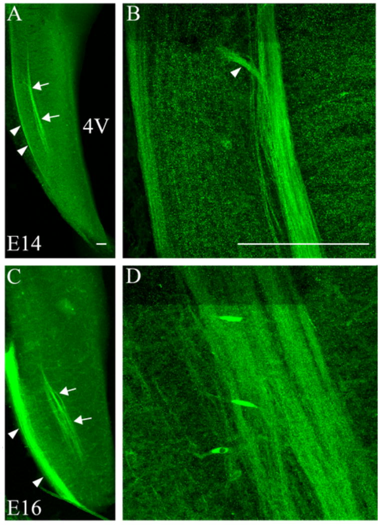
Figure 2. Neuropilin-1 is expressed in the ST and trigeminal tract in embryonic brainstem.

Neuropilin-1 expressing fibers were observed in the ST at E14 and E16. A. At E14, a low magnification image shows Npn-1 positive fibers in the ST (arrows) and in the more lateral trigeminal tract (arrowheads). B. At higher magnification, clear fasciculation of the ST fibers is apparent (arrowhead indicates entering ST fibers). C. The same pattern of Npn-1 expression in the ST is observed at E16 in a low magnification image of the brainstem (arrows indicate ST; arrowheads indicate trigeminal tract). D. The higher magnification image illustrates fasciculation of the ST fibers at E16. 4V = 4th ventricle. Scale bars = 100 μm.
