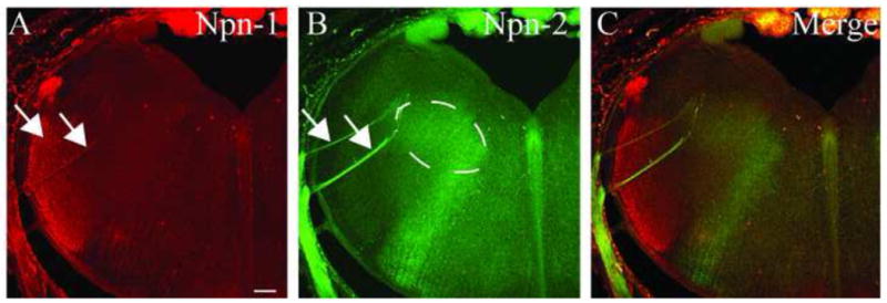
Figure 5. Neuropilin-1 and Neuropilin-2 expression in the coronal plane.

A, B. E16 coronal brainstem sections illustrating expression patterns of Npn-1 (A) and Npn-2 (B). The branches of cranial nerve VII are highlighted by arrows. The dashed line in B indicates the area of the Npn-2-labeled, tuft-like structures. C. A merged image details the distinct populations of tuft-like structures and trigeminal fibers, whereas the VII nerve projections express both Npn-1 and Npn-2. Scale bar = 100 μm.
