
Fig. 1.
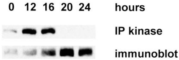
RING3 kinase activity and protein level after serum stimulation of fibroblasts. Extracts were prepared at progressive times from 107 serum-stimulated Swiss/3T3 cells and immunoprecipitated with αRING3 rabbit polyclonal antibody. Immune complexes were separated by SDS-PAGE, electroblotted to PVDF, and subjected to autophosphorylation assay (Ref. 1; IP kinase) or αRING3 immunoblot.