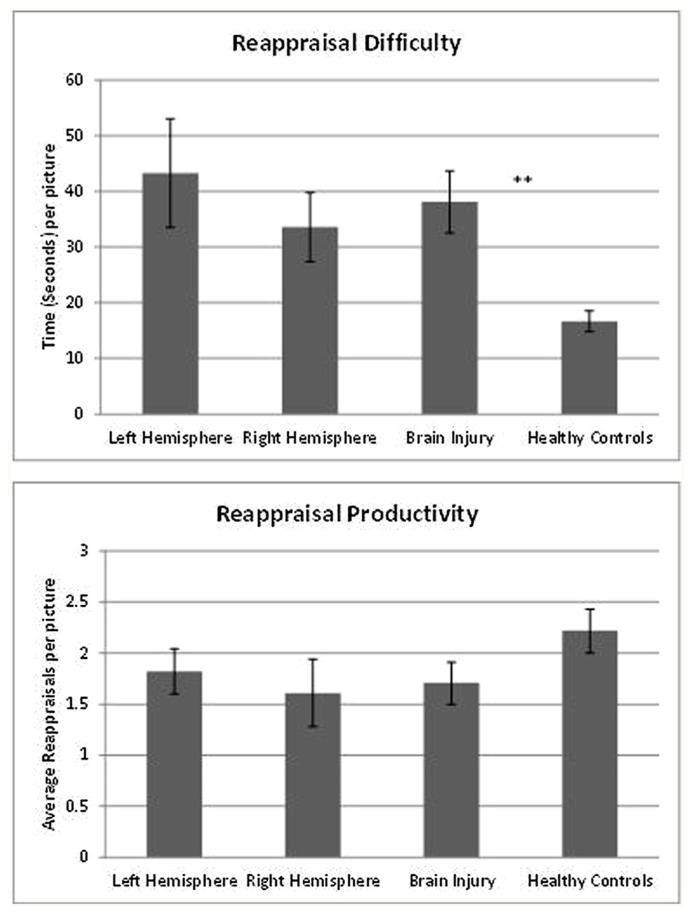
FIGURE 1.
Laterality and reappraisal generation. The figures describe differences between neurological groups and controls in reappraisal difficulty and productivity. Patients with left hemisphere and right hemisphere lesions are equally slow generating the first reappraisal (top figure). However, they do not differ from controls in the average number of reappraisal produced in each picture (bottom figure), **p < 0.001.

