
Figure 1.
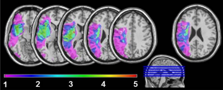
Lesion sites overlapping in 1, 2, 3, 4, or 5 of the agrammatic speakers. For the very right axial slice, first a lesion overlay of 11 non-agrammatic aphasic speakers was plotted. Next the brain areas, where at least 6 lesions overlapped, were subtracted from the overlay plot of lesion sites in the five agrammatic speakers to adjust for areas with higher anatomical vulnerability.