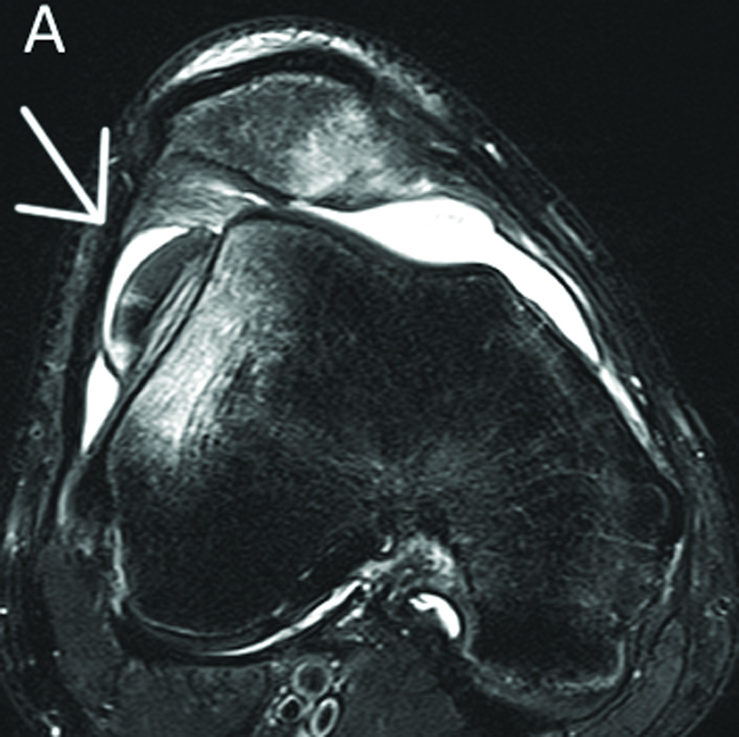
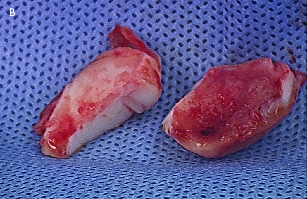
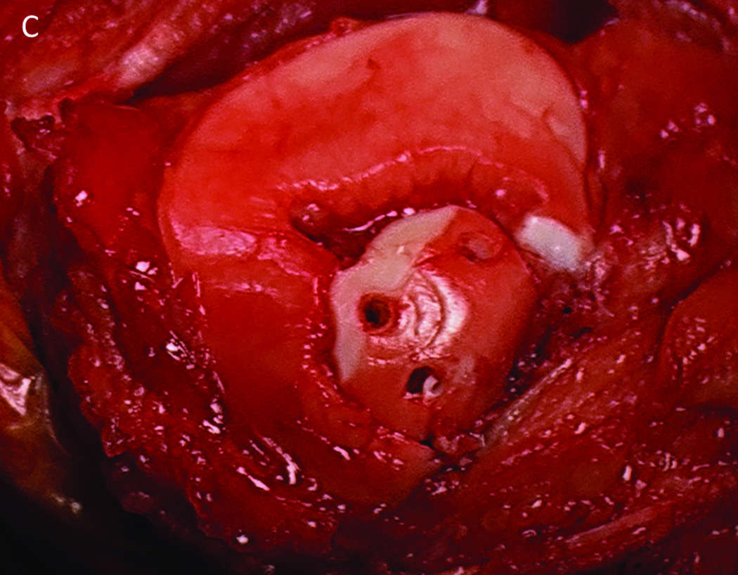
Figure 3.
MRI after an acute high-energy patellar dislocation. The arrow indicates a large osteochondral body from the medial facet of the patella (a). This fragment was excised and one portion (right side) was found to have bone attached while one section (left side) did not (b). This fragment was affixed to the medial side of the patella with biocompression screws (c).


