Figure 2.
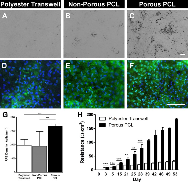
fhRPE cultured on porous PCL display improved markers of maturity including pigmentation, improved tight junction localization, increased cellular density, and superior barrier formation. Light microscopy images of (A) porous polyester transwell, (B) nonporous PCL, and (C) porous PCL displaying varying levels of fhRPE pigmentation at 8 weeks. Immunohistochemical staining for tight junction associated protein-1 (ZO-1, green) and nuclei (DAPI, blue) in cells cultured for 8 weeks on (D) porous polyester transwells, (E) nonporous PCL, and (F) porous PCL. Scale bars: 100 μm. (G) Graph indicating significantly increased RPE cellular density on porous PCL compared to either control substrate after 8 weeks. (H) Increased transepithelial resistance of fhRPE on porous PCL throughout the first 8 weeks of culture indicating improved barrier formation. Please note that data from >28 days is derived from only two biological replicates, so no statistical measures were used. **P < 0.01; ***P < 0.001.

