Fig. 1.

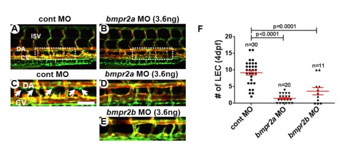
Reduction in Bmpr2a and 2b activity causes loss of lymphatic endothelial cells in thoracic duct of zebrafish. Confocal images taken from the trunk region of 4dpf control (A, C), bmpr2a (B, D), and bmpr2b (C) MO-injected embryos in Tg(fli1a:negfp); Tg(kdrl: mCherry) double transgenic background. GFP+ mCherry− cells are the lymphatic endothelial cells (LECs) within the thoracic duct (white arrows). (F) Quantification on the number of LECs in control, bmpr2a, and bmpr2b MO-injected embryos. LECs in the TD between 8th and 15th somite were quantitated by confocal imaging analyses. Areas within the white rectangles in (A) and (B) are shown in higher magnification in (C) and (D). DA, dorsal aorta; CV, cardinal vein; ISV, intersegmental vessel. Scale bar is 50 μm.
