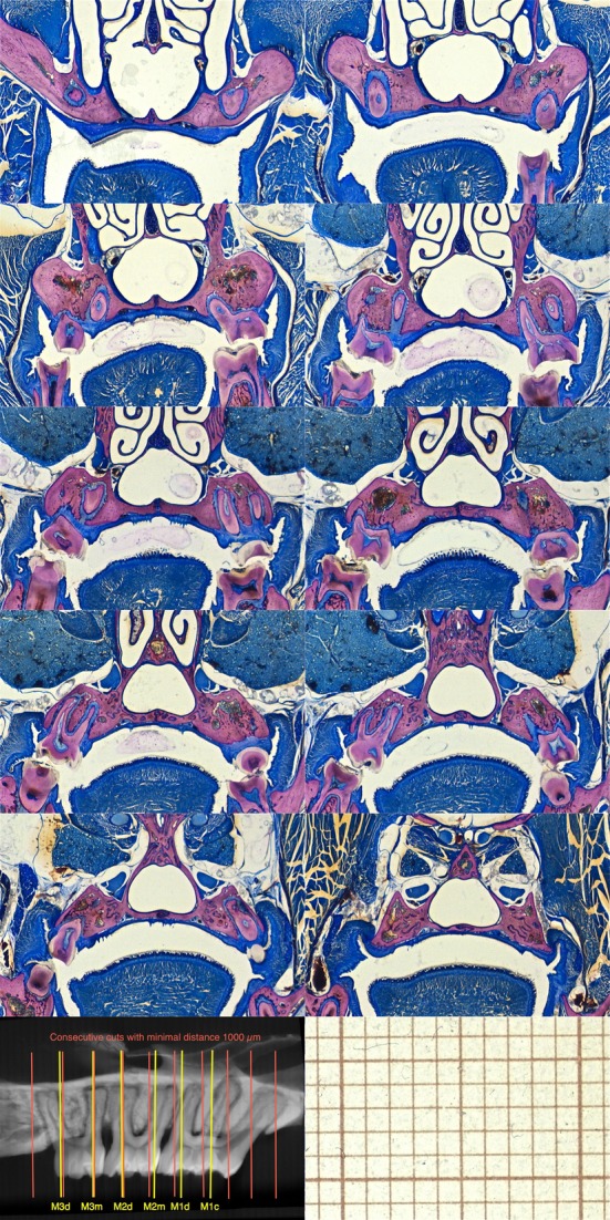
Figure 1.

Consecutive histomicrographs every 1000 μm (red lines) of an undissected control rat. Some of the buccal roots were located between the cutting planes and thus do not appear on the sections. In most of the cases, it is not possible to evaluate both sides on the same section. MPR-aligned virtual slices located at lateral root prominences normally include the pulp chamber at the buccal bone level (M1c, M1d, M2m, M2d, M3m, M3d). At these sites dental parameters were evaluated (yellow lines).
