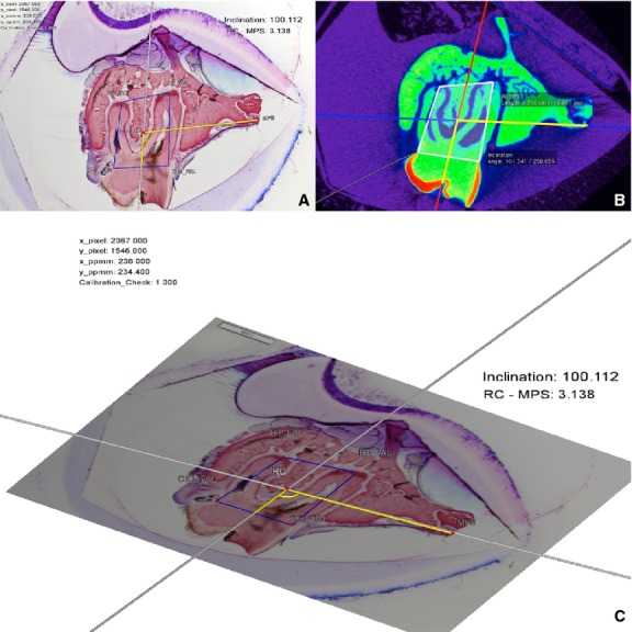
Figure 4.

MPR-aligned virtual micro-CT section (A) and the corresponding histomicrograph of the obtained section (B). The same measurements made on micro-CT and histomicrographs were compared to evaluate methodological errors and differences between the methods regarding tooth position and inclination. (C) 3D display of the interactive geometrical construction by the software DreiDEdit Archimedes Geo 3D.
