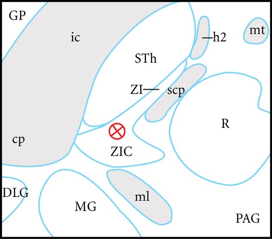
Figure 6.

Scale diagram of an example of a modern clinical posterior subthalamic area deep brain stimulation (DBS) target 3.5 mm ventral to the modified axial commissural plane of Schaltenbrand & Wahren (1977). This plane is favoured by neurosurgeons for DBS planning. The target shown (a red circle and cross) represents an attempt to optimise neuromodulation of the caudal zona incerta (ZIC) and posterodorsal subthalamic nucleus (STh). In this position, the effects of proximity to the internal capsule (ic), medial lemniscus (ML) and the ventromedial subthalamic nucleus are minimised. The result is a reduction in clinical side effects such as skeletal muscle contraction, dysaesthesiae, and limbic effects, respectively. The combination of surgical targeting error and the relatively symmetrical current spread from the electrode is a source of side effects. Moreover, these factors create uncertainty as to which brain structures underlie positive clinical effects in parkinsonism and tremor. This composite diagram has been adapted from drawings of human brain horizontal sections as shown in diagram LXVIII (−3.5) of Schaltenbrand & Wahren (1977) and horizontal sections V2.7 and V3.6 of Morel (2007). In this diagram, the midline is to the left, and rostral is at the top. Other structures shown in this diagram are the central part of the zona incerta (ZI), red nucleus (R), medial geniculate (MG), cerebral peduncle, dorsal lateral geniculate nucleus (DLG), superior cerebellar peduncle (scp), lenticular fasciculus (h2), mammilothalamic tract (mt), globus pallidus (GP), and periaqueductal grey (PAG).
