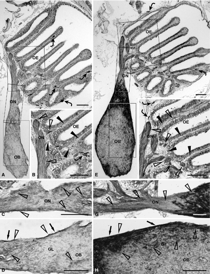
Figure 1.

Immunohistochemical detection of olfactory ensheathing cell markers, GFAP (A–D) and S100 (E–H), in the olfactory system of zebrafish. (A) Distribution of GFAP immunopositivity in the complete olfactory system. Rectangular areas marked with OE, ON and OB are shown in (B), (C) and (D), respectively, at higher magnification. Some chromatophores (curved arrows) are present in the epithelial lamina propria. (B) GFAP immunopositivity (open arrowheads) appears slight in the epithelial lamina propria (arrowheads). A chromatophore (curved arrow) is visible. (C) The olfactory nerve shows clear GFAP immunopositivity (open arrowheads). (D) In the olfactory bulb, GFAP immunostaining (open arrowheads) is more evident in the olfactory nerve layer (arrows) than in the glomerular layer. (E) Distribution of S100 immunostaining in the whole olfactory system. Rectangular areas marked with OE, ON and OB are shown in (F), (G) and (H), respectively, at higher magnification. Some chromatophores (curved arrows) are evident in the epithelial lamina propria. (F) The epithelial lamina propria (arrowheads) shows clear immunostaining (open arrowheads) and some chromatophores (curved arrows). (G) In the olfactory nerve, the intracranial tract is heavily stained, compared with the extracranial portion (open arrowheads). (H) In the olfactory bulb, the olfactory nerve layer (arrows) exhibits intense staining (open arrowheads). Evident immunostaining (open arrowheads) appears in the glomerular layer. GL, glomerular layer; OB, olfactory bulb; OE, olfactory epithelium; ON, olfactory nerve. Scale bars: 100 μm.
