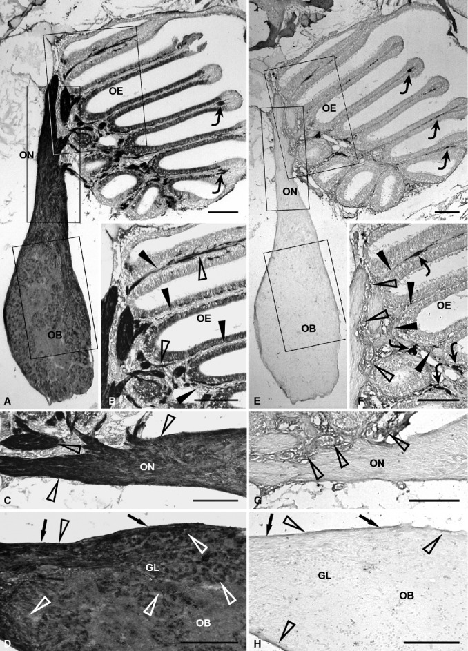
Figure 2.

Immunohistochemical detection of olfactory ensheathing cell markers, NCAM (A-D) and PSA-NCAM (E-H), in the olfactory system of zebrafish. (A) Evident NCAM staining in the whole olfactory pathway. Rectangular areas marked with OE, ON and OB are shown in (B), (C) and (D), respectively, at higher magnification. The epithelial lamina propria contains some chromatophores (curved arrows) (B) The epithelial lamina propria (arrowheads) shows moderate immunostaining (open arrowheads). (C) The extracranial olfactory nerve (open arrowheads) shows stronger expression compared with the intracranial olfactory nerve. (D) In the olfactory bulb, the olfactory nerve layer (arrows) and glomerular layer are clearly immunostained (open arrowheads). (E) In the olfactory pathway, PSA-NCAM immunostaining appears weak. Rectangular areas marked with OE, ON and OB are shown in (F), (G) and (H), respectively, at higher magnification. Some chromatophores (curved arrows) are present in the epithelial lamina propria. (F) Weak PSA-NCAM immunoreactivity (open arrowheads) in the lamina propria (arrowheads) of the olfactory mucosa. Curved arrows point out some chromatophores. (G) The olfactory nerve is unstained. Immunostaining (open arrowheads) appears in the epithelial lamina propria. (H) In the olfactory bulb, weak immunopositivity (open arrowheads) is observed in the olfactory nerve layer (arrows), while the glomerular layer is unstained. GL, glomerular layer; OB, olfactory bulb; OE, olfactory epithelium; ON, olfactory nerve. Scale bars: 100 μm.
