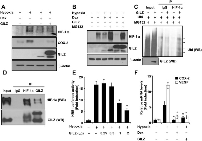
Figure 4.
GILZ physically interacts with HIF-1α and serves as a negative regulator of HIF-1. (A,B) A549 cells were transfected with pcDNA-GILZ expression plasmids. At 24 h post-transfection, cells were pretreated with dexamethasone (0.1 μM) for 1 h before treatment with hypoxia for 24 h and analysed by Western blot (WB). (C) A549 cells were transfected with Ubi or pcDNA-GILZ plasmid as indicated. At 36 h post-transfection, cells were treated with 10 μM MG132 for 12 h. Ubi-conjugated HIF-1 was detected using anti-ubiquitin antibody. Arrows indicate the ubiquitinated HIF-1 protein bands. (D) After transfection with pcDNA-GILZ and HIF-1α, the whole cell lysates were immunoprecipitated (IP) with GILZ or HIF-1α antibody, and WB was performed with GILZ or HIF-1α antibody after immunoprecipitation. The expression of proteins was analysed by WB using GILZ or HIF-1α antibody as input. (E) A549 cells were transfected with HRE-Luc reporter with or without pcDNA-GILZ expression plasmids, treated as indicated, and luciferase activity was assayed. (F) A549 cells were transfected with pcDNA-GILZ expression plasmids and treated as indicated. COX-2 and VEGF mRNA expression was quantified by qPCR. Values represent the mean ± SD (n = 3). *P < 0.05. All experiments were repeated at least three times.

