
Figure 7. NUAK1 inhibition suppresses cell proliferation.
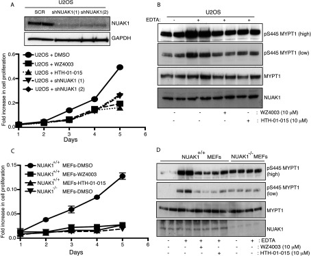
(A) U2OS cells were incubated with or without 10 μM WZ4003 or 10 μM HTH-01-015 and a cell proliferation assay was carried out over 5 days in triplicate using the CellTiter 96® AQueous Non-Radioactive Cell Proliferation Assay kit (Promega) (as described in the Materials and methods section). U2OS cells in which NUAK1 has been knocked-down using two different shRNA hairpins were used in parallel as controls. The efficiency of the knock down of each shRNA is shown in top panel. SCR, control scrambled shRNA hairpin; shNUAK1 (1), first NUAK1 shRNA hairpin; shNUAK1 (2), second NUAK1 shRNA hairpin. (B) U2OS cells were treated with (+) or without (−) 10 μM WZ4003 or 10 μM HTH-01-015. After 16 h cell media was removed and cells were treated with EDTA-PBS-based cell dissociation buffer supplemented with 10 μM WZ4003, 10 μM HTH-01–015 or DMSO for 20 min. Cell detachment was induced with gentle tapping of the plates followed by gentle centrifugation at 70 g for 3 min. Cells were lysed immediately after removal of the media and immunoblotted for the detection of the indicated antibodies. (C and D) As above, except NUAK1+/+ and NUAK1−/− MEFs were used. Similar results were obtained in three separate experiments.