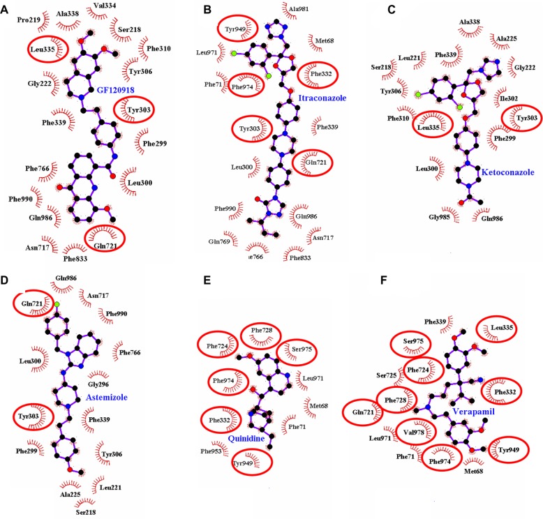
Figure 5.
Receptor–ligand interaction diagrams for six binding sites of the same protein (murine 3G60 P-glycoprotein) each with a different ligand molecule bond. The ligands are: (A) GF120918, (B) itraconazole, (C) ketoconazole, (D) astemizole, (E) quinidine, and (F) verapamil. The spoked arcs represent protein residues making nonbonded (hydrophobic) contacts with the ligand. The contacted ligand atoms are shown with spokes radiating back. The red ellipses in each plot indicate protein residues that are in common to those associated with the QZ59-RRR ligand.
Abbreviation: QZ59-RRR, cyclic-tris-(R)-valineselenazole.

