
Fig. 1.
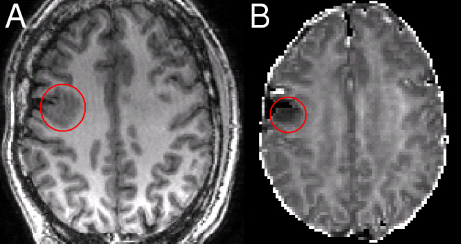
Residual FCD following a right middle frontal gyrus resection. Patient 1: the residual FCD on the T1-weighted image (A) corresponds with an area of reduced ICVF (B).
Official websites use .gov
A
.gov website belongs to an official
government organization in the United States.
Secure .gov websites use HTTPS
A lock (
) or https:// means you've safely
connected to the .gov website. Share sensitive
information only on official, secure websites.
Residual FCD following a right middle frontal gyrus resection. Patient 1: the residual FCD on the T1-weighted image (A) corresponds with an area of reduced ICVF (B).