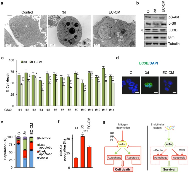
Figure 4. Brain endothelial cells protect GSC against mitogen deprivation-induced autophagy and apoptosis.
a-d) GSC#1 were cultured for 3 days with control medium (C), deprivation medium (3d) or endothelial-derived conditioned medium (EC-CM). a) Electron microscopy analysis revealed a protective effect of EC-CM against apoptosis when compared to 3 day-starved cells. Scale bars: 1 μm. b) Protein extracts were analyzed by western blot for the indicated antibodies. ** non-processed form; * processed form. c) GSCs #1–14 were cultured for 3 days with deprivation medium or EC-CM and PI incorporation was measured by flow cytometry. Percent of cell death was represented as the mean+s.d. of three independent experiments. Student’s t-test: *** P<0.001, ** P<0.01, * P<0.05. d) LC3B puncta was examined by confocal microscopy. Scale bars: 5 μm. e-f) Annexin V-FITC/PI staining (e), as well as PI staining alone (f), were used to evaluate apoptosis as described in 1f-g. Each panel is representative of three independent experiments. g) Schematic representation of the central role of mTor in regulating mitogen deprivation-induced autophagy and apoptosis in GSCs (left panel). The positive effect of endothelial factors on this pathway is illustrated (right panel).

