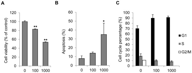
Figure 1. IFN-γ decreased viability of melanocytes, caused apoptosis and cell cycle arrest.

Primary normal human Melanocytes were treated with various concentrations of IFN-γ (0, 100 or 1000 U/ml) for 72 h. Cell viability was then examined by MTS assay (A). Apoptosis was analyzed by flow cytometry after cells were stained with PI and Annexin V-FITC (B). (C) Cell cycle distribution of melanocytes was measured 24 h post IFN-γ treatment. Results are presented as mean ± SD from at least three independent melanocyte cultures. *P<0.05, **P<0.01, Student's t-test compared with controls.
