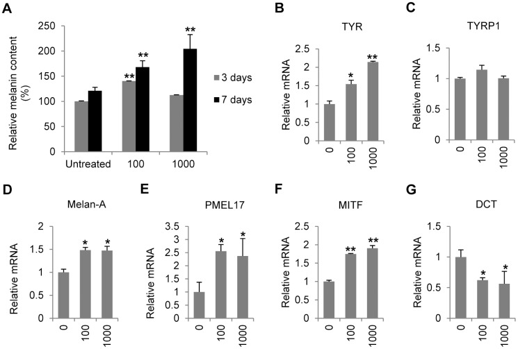
Figure 2. Effects of IFN-γ on melanogenesis in normal melanocytes.
(A) Melanocytes were treated with various concentrations of IFN-γ (0, 100 or 1000 U/ml) for 3 or 7days before melanin content was measured. The melanin content was normalized on the basis of protein concentration. (b–g) Total RNA was extracted from melanocytes treated with or without IFN-γ for 24 hours. Real-time PCR was then performed to evaluate the relative mRNA levels of (B) tyrosinase (TYR), (C) tyrosinase-related protein 1 (TYRP1), (D) Melan-A, (E) melanocyte protein 17 (PMEL17), (F) microphthalmia-associated transcription factor (MITF), and (G) dopachrome tautomerase (DCT). The values shown represent the mean ± SD of three independent melanocyte cultures. *P<0.05 and **P<0.01.

