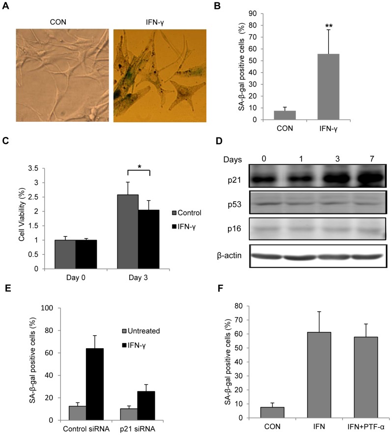
Figure 3. IFN-γ caused senescence in melanocytes through p21 pathway.
Melanocytes were treated with or without 100/ml IFN-γ for 7 days. Senescence was evaluated based on SA-β-gal staining, and cell morphology. (A) Representative pictures of SA-β-gal-stained cells observed under bright-field microscope. Flattened and enlarged cells with blue/green stain were regarded as senescent cells (B) Quantification of SA-β-gal-positive cells based on microscopic analysis. CON represents the control cells. **P<0.01. (C) After 7 days of treatment, melanocytes were cultured in fresh medium without IFN-γ for 3 days and cell viability was examined by MTS assay. (D) Melanocytes were cultured in the presence or absence of IFN-γ for up to 7 days and cells were harvest on day 1, 3 and 7. Cell lysates were subjected to SDS-PAGE and analyzed by western blot with indicated antibodies. β-actin was probed as the loading control. (E,) Bar graphs of SA-β-gal staining results. (E) Melanocytes were transfected with scrambled control or p21 siRNAs for 48 h before IFN-γ treatment. (F) Melanocytes were treated with or without 100 U/ml IFN-γ for 7 days in the presence of DMSO or 20 µM pifithrin-α (PFT-α).

