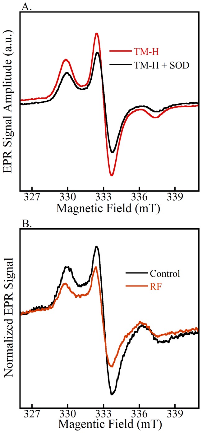
Figure 7. EPR spectra of cyclic hydroxylamines in cell free controls and an example of RF experiments for the detection of superoxide.
(A) PEG-SOD (50 U/ml) inhibits the EPR signal by up to 40% in a cell-free xanthine/xanthine oxidase system. (B) Control and RF normalized EPR spectra. TM-H spin probe reacts with intracellular superoxide to give a nitroxide free-radical that is detectable by EPR. The RF samples have a lower EPR signal intensity compared to control, indicative of a lower intercellular superoxide concentration.

