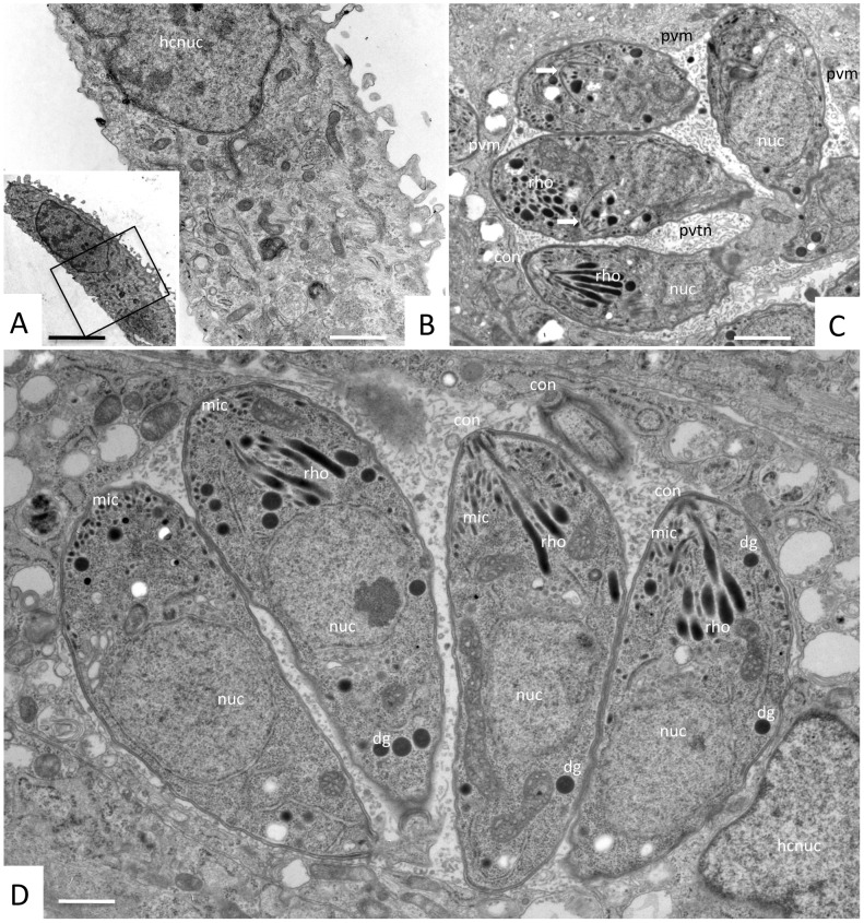
Figure 4. TEM analysis of compound 1294 treated and untreated N. caninum-infected HFF cultures at 3 days post-infection.
A shows a HFF cell with inhibitor 1294 (2. 5 μM) added at the time point of infection, B represents a higher magnification view of A. Note the absence of parasites, and the fact that compound 1294 does not induce any obvious ultrastructural alterations within the host cell. C and D show N. caninum-infected HFF cultured in the absence of the inhibitor at 3 days post-invasion. Numerous tachyzoites are located within parasitophorous vacuoles, surrounded by a parasitophorous vacuole membrane (PVM), and embedded in a matrix of a tubular membrane network (PVTN). Apical parts of tachyzoites are characterized by the conoid (con), micronemes (mic), and rhoptries (rho); dg = dense granules, nuc = tachyzoite nucleus, hcnuc = host cell nucleus. The two arrows in C point towards proliferating tachyzoites undergoing endodyogeny. Bars in A = 5 μm; B = 1 μm; C = 0.58 μm; D = 0.28 μm.

