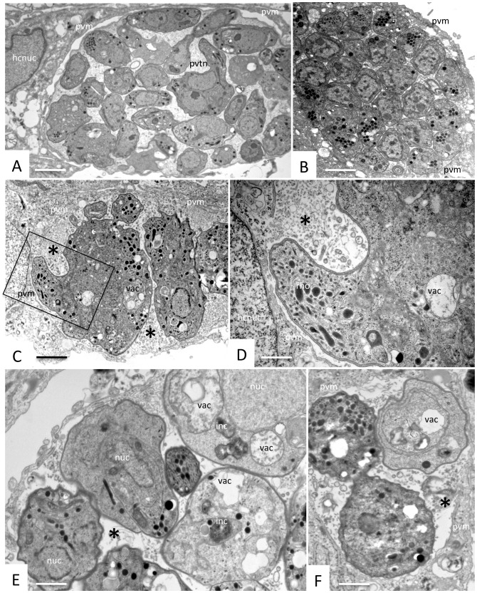
Figure 5. TEM analysis of N. caninum-infected HFF cultures treated for 3 days, with 2.5 μM of inhibitor 1294 added at 2 h post-infection.
A and B show micrographs of more or less densely packed parasitophorous vacuoles containing numerous tachyzoites without obvious alterations. C and D show a representative example of a vacuole delineated by a parasitophorous vacuole membrane (pvm) containing parasites displaying a large cytoplasmic mass and aberrant overall morphology. The boxed area in C is enlarged in D, exhibiting the presence of the pvm and rhoptry-like organelles (rho). In many instances, as seen in Figure 5E and F, parasitophorous vacuoles contain several parasites exhibiting clear signs of metabolic impairment such as cytoplasmic vacuolization (vac) and electron-dense inclusions (inc). (Figure 5E, F). Note that in C–F the matrix has lost its characteristic tubular network structure and is now formed of either granular material or possibly membranous material (C, D), or is even largely missing (E. F). Bars in A = 1 μm; B = 0.9 μm; C = 0.75 μm, D = 0.35 μm; E = 0.3 μm; F = 0.3 μm.

