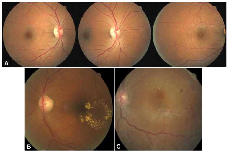
Figure 1.
Images obtained according to the EyePACS imaging protocol. (A) normal retina: left primary field OD-temporal retina and optic nerve, center) field centered on optic nerve OD, right) optic nerve and nasal retina OD. (B) and (C) would each yield referrals for retinal edema because of hard exudates within 1 disc diameter of foveola. (B) circinate ring of hard exudates involving macula OS,(C) small hard exudates within1 disc diameter of foveola.w

