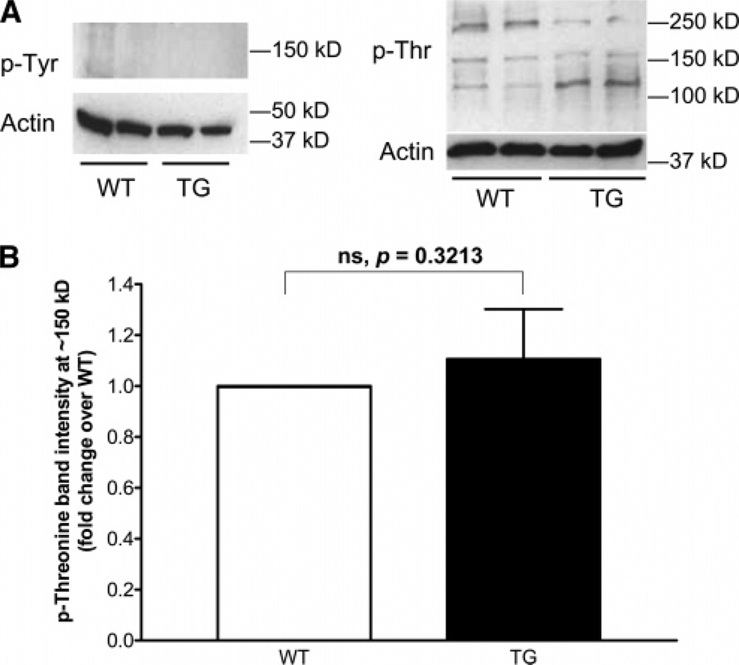
Figure 2.
Western blotting of the ventricular myofibrillar proteins extracted from 12-month-old WT or TG mouse hearts for Tyr or Thr phosphorylation using the p-Tyr antibody or the p-Thr antibody. Five micrograms of sample was loaded in each lane; Western blotting for actin was used as control for loading. (A) Representative results of Western blotting for Tyr and Thr phosphorylation. (B) Quantitative densitometry analysis of the Western blot results using p-Thr antibody; n = 3 (from three pairs of hearts). The density of the p-Thr at ~150 kDa was normalized to that of the stripped and reprobed actin in the same sample. The data were then presented as fold change in Thr phosphorylation over that of the WT hearts. The paired, one-tailed t-test was used for statistical analysis. p = 0.3213; ns, not significant.

