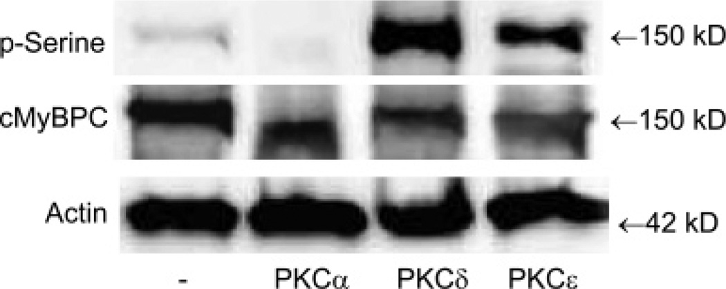
Figure 7.
In vitro PKC assay on purified cardiac myofibrillar proteins from 12-month-old WT mice followed by Western blotting. A representative Western blot result of three independent experiments was shown. Five micrograms of sample was incubated with or without an equal amount of PKCα, PKCδ, or PKCε at 30 °C for 20 min. In order to activate PKCα, the PKCα assay buffer contained 200 µM CaCl2. The p-Ser and actin were first detected by Western blotting. The membrane was then stripped and reprobed for cMyBPC.

