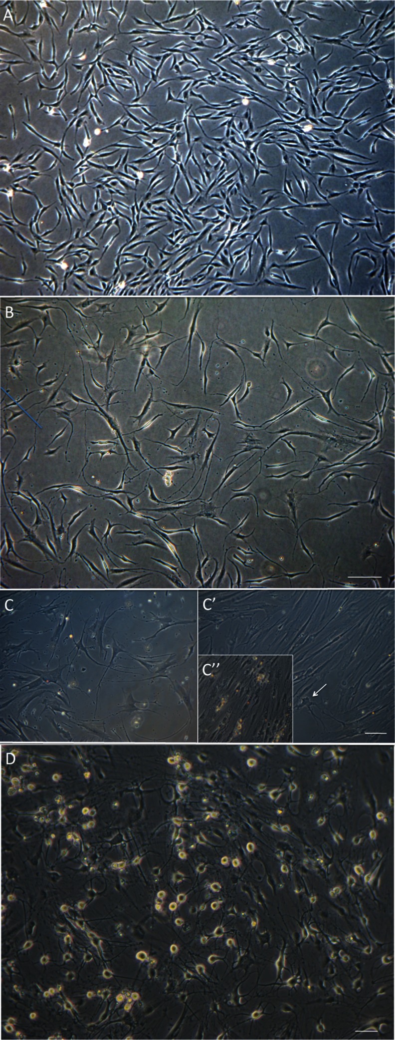
Fig. 1.
Phase contrast images of differentiating hEPI-NCSC. a hEPI-NCSC with stellate morphology in expansion culture prior to differentiation. b By differentiation day 4, virtually all cells were bipolar with short processes. Bar, 100 μm. c In the presence of IWP-4 virtually all cells were multipolar with long processes and dopaminergic neuron morphology. In contrast, in the absence of IWP-4, the majority of cells often, but not always, flattened and lost neuronal morphology by day 14–18 (C’). Many neurons appeared unhealthy (e.g., arrow) and subsequently died. In subsequent days there were areas with widespread cell death (C”). Bar, 100 μm. d By day 25–30 differentiated cells had assumed mature multipolar neuronal morphology with phase-bright somata. Bar, 50 μm

