Fig. 4.
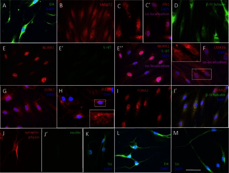
Immunocytochemistry at day 25 in culture. a All cells expressed dopamine (DA); neuronal morphology is evident. b All cells were immunoreactive for the vesicular transporter VMAT2. c EN1 was expressed in all nuclei (inset) and also in perinuclear areas. d All cells expressed the early neuronal marker, β-III tubulin. (E-E”) NURR1/serotonin double stain. All cells expressed intense nuclear NURR1 immunofluorescence (e, E”); (E’) background serotonin (5HT) immunoreactivity; (E”) merged E and E’ images with blue DAPI nuclear stain. f LMX1b was expressed in all nuclei (inset). There was also unspecific cytoplasmic fluorescence. g GIRK2 was expressed in all cells (GIRK2/DAPI merged image). h PITX3 in 91.6 ± 14.6 % of cells; there was unspecific cytoplasmic fluorescence that interfered with image acquisition; inset shows higher magnification of a nucleus. i FOXA2 was localized in the nucleus of 89.6 ± 19.4 % of cells. j Synaptophysin/nestin double stain; (J’) all cells were intensely synaptophysin immunoreactive whereas (J”) nestin immunoreactivity was at background levels. k, l, m All cells expressed TH in the soma and in processes. Bar, (a–m), 50 μm

