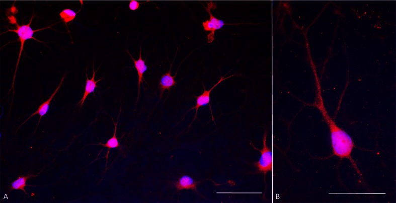
Fig. 5.
Dopamine immunocytochemistry at day 30 of culture. a Double stain with antibodies against dopamine (red fluorescence) and DAPI nuclear stain (blue fluorescence) shows that all cells express dopamine and have mature dopaminergic neuronal morphology. Contrast is exaggerated in order to visualize fine processes. b Typical dopaminergic neuron morphology at higher magnification showing punctate, presumably vesicular, dopamine immunoreactivity

