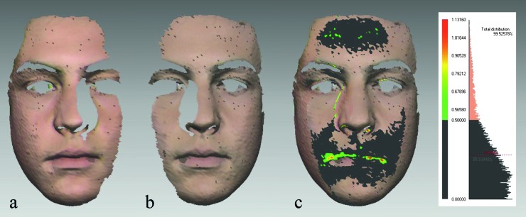
Figure 1.
Evaluation of laser scan’s quality. (a and b) Right and left facial halves (facial shells) in Rapidform 2006® (INUS Technology, Seoul, Korea) after four-stage processing. (c) Absolute colour map and the histogram were used to evaluate scan quality before merging. Surface matching of the two shells in the overlap area was 85.53 per cent. Deviations less than 0.5 mm are presented in dark grey, 0.51–0.79 mm in light green, 0.80–0.90 mm in yellow, and 0.91–1.13 mm in red. Internally developed subroutine automatically determined average distance between the facial shells in the overlap area: 0.28 mm (SD 0.24 mm). Therefore, laser scans were suitable for merging.

