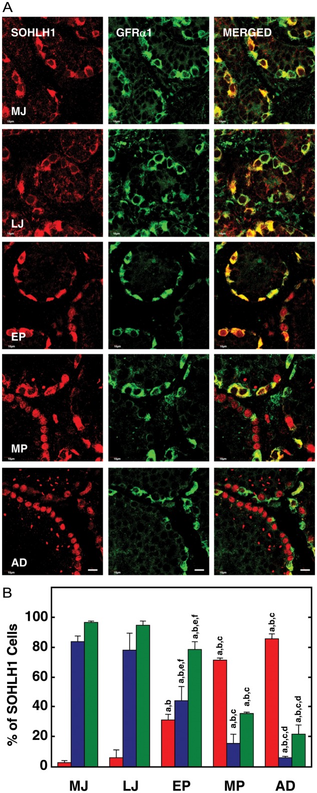
Figure 2.

(A) Confocal projections (100×; 1 µm optical sections) illustrating the distribution of SOHLH1 (left-hand panels) and GFRα1 (middle panels) immunostaining in 5 µm sections of testis from a MJ, LJ, EP, MP and AD rhesus monkey. The merged image of the two signals is shown in the right-hand panel. Note the predominantly cytoplasmic staining for SOHLH1 in the juvenile testis and that it is co-localized with that for GFRα1. With the initiation of puberty, SOHLH1 begins to exhibit a nuclear location which is exemplified in the adult. Scale bar, 10 µm. (B) The percentage (mean ± SEM) of immunopositive SOHLH1 spermatogonia exhibiting nuclear-only staining (red bars), cytoplasmic-only staining (blue bars) and co-expressing GFRα1 (green bars) in testes from MJ, LJ, EP, MP and AD rhesus monkeys. Note the progressive and dramatic increase in nuclear location of SOHLH1 that is initiated with the onset of puberty and is associated with a reduction in GFRα1 staining. n=3 for each developmental group. Letters on top of the bars denote significant differences from MJ (a), LJ (b), EP (c), MP (d and e) and AD (f).
