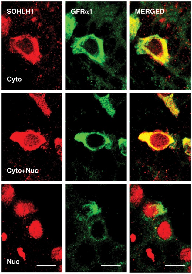
Figure 3.

High-power confocal projections of dual immunofluorescence labeling of SOHLH1 (left hand panels) and GFRα1 (middle panels) in 5 µm sections of monkey testis. The right-hand panels show the merged signal. Three patterns of SOHLH1 staining of spermatogonia staining were observed: cytoplasmic-only (top panels), cytoplasmic and nuclear (middle panels) and nuclear-only (lower panels). Scale bar, 10 µm.
