
Fig. 2.
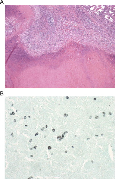
Thoracoscopic lung biopsy of the left upper lobe nodule demonstrates a necrotizing granuloma containing pneumocystis organisms surrounded by a hyal- inized capsule. A. Section of necrotizing granuloma with necrotic centre. Organisms are not visible on this stain. Hematoxylin and eosin stain at 40× magnification B. Organisms with the typical size and shape of Pneumocystis can be seen outlined in black. Grocott's Methenamine Silver stain at 1000× magnification.