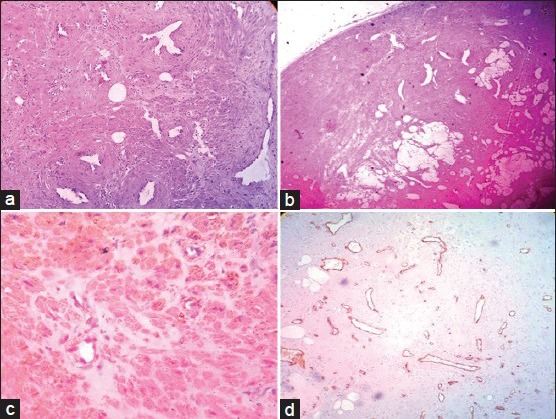
Figure 1.

(a) Inner layer of muscle are arranged circumferentially around vessels and outer layer blends with less well-ordered smooth muscle tissue of tumor. (H and E, ×40) (b) Islands of lipometaplasia within the tumor. (H and E, ×40) (c) Diffuse cytoplasmic positivity present in tumor cells for smooth muscle actin. (×100) (d) CD34 positivity seen only in endothelial cell of vessel wall. (×40)
