
Figure 4.
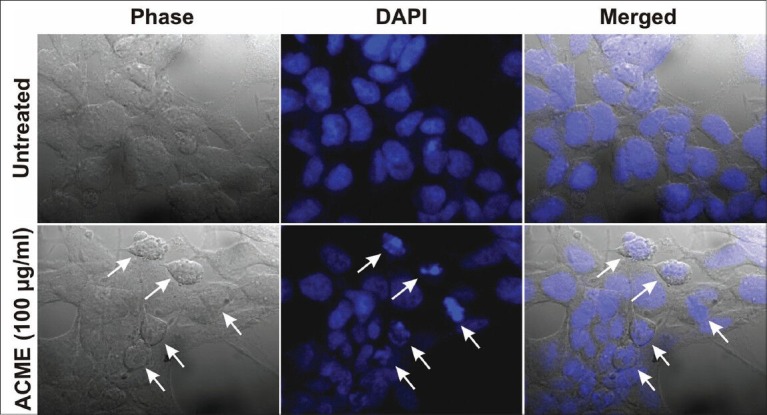
Morphological assessment of ACME treated MCF-7 cells for 48 hours. Nuclei were stained with DAPI and observed under confocal microscope. Untreated MCF-7 cells (upper region) and MCF-7 cells treated with 100μg/ml ACME (lower region). White arrows indicate the cells which have undergone apoptosis