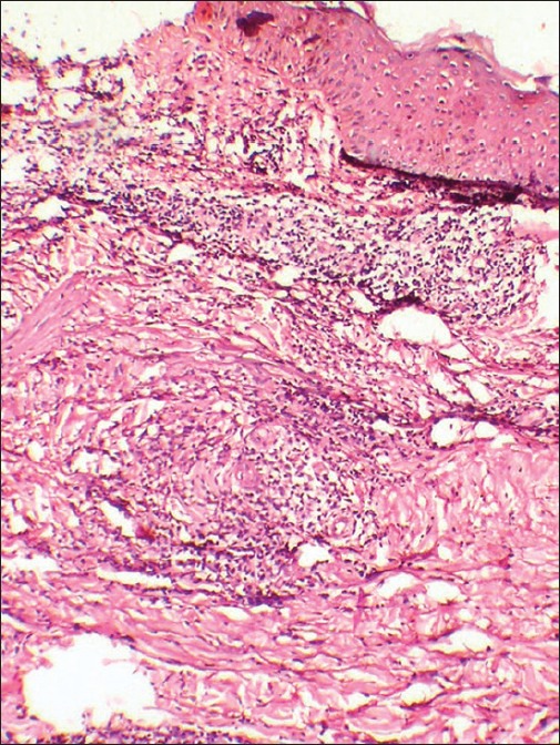
Figure 4.

Histopathology of skin lesions revealing prominent upper dermal edema with a heavy neutrophilic infiltrate in the upper dermis and a coalescing infiltrate of neutrophils in the lower dermis especially around blood vessels with absence of vasculitis (H and E, ×10)
