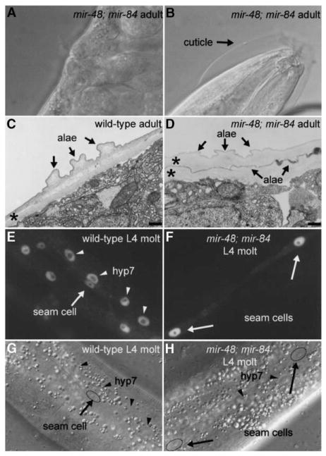
Figure 2. mir-48; mir-84 Double Mutants Display Supernumerary Molting Behavior in Adult Worms.
(A and B) Nomarski DIC images of a mir-48; mir-84 animal with a (A) fully formed vulva and with embryos visible inside of the worm to show that the worm is in the adult stage and (B) unshed cuticle surrounding the anterior region of the worm.
(C and D) Electron micrographs of adult-stage cuticle in a (C) wild-type animal and a (D) mir-48; mir-84 adult. Asterisks indicate cuticles. The scale bar is 300 nm. (C) Normal single cuticle with alae in a wild-type adult. (D) Two cuticles, both with alae structures (arrows), are visible in a mir-48; mir-84 adult.
(E and F) Fluorescence micrographs of a (E) wild-type and a (F) mir-48; mir-84 worm at the L4 molt stage that carry the col-19::gfp transgene maIs105. (E) Fluorescence micrograph of a wild-type L4 molt-stage worm. Expression of col-19::gfp is observed in nuclei of the hyp7 syncytium (arrowheads) and hypodermal seam cells (arrows) in a wild-type L4 molt-stage-worm. (F) Expression of col-19::gfp is observed in hypodermal seam cells (arrows) in a mir-48; mir-84 L4 molt-stage worm. col-19::gfp expression is reduced or absent in hyp7.
(G and H) Nomarski DIC images of the hypodermis of the animals in (E) and (F), respectively, showing seam cell (arrows) and hyp7 (arrowheads) nuclei.

