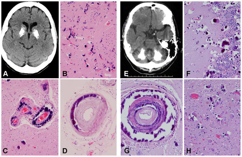
Figure 1. Novel pathogenic SLC20A2 mutations: clinical and pathological manifestations.
A-D (p.S113*): Brain axial unenhanced computed tomography image of the p.S113* carrier shows marked symmetric calcification of the basal ganglia (A). Histologic studies showed extensive small vessel calcific vasculopathy and calcospherites in the putamen (B); large and small vessel deposits in the globus pallidus (C) and deposits in arteries in subarachnoid space (D). E–H (SLC20A2 genomic deletion): Brain axial unenhanced computed tomography image of pedigree member II-3 shows marked extensive bilateral cerebellar calcifications (E). Histologic studies showed extensive small vessel calcific vasculopathy and many calcospherites in the cerebellar molecular and internal granular cell layer (F); intramural deposits in large vessels in the cerebellar deep white matter (G); and calcific deposits in the deep cerebellar nuclei (H).

