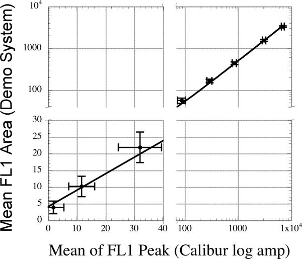
Figure 7.
A linear fit of the fluorescence response of the peak values collected in logmode from the native FACSCalibur data acquisition system vs. the fluorescence area values collected by the demonstration system. The fit was weighted by the inverse of the square of the standard deviation of each point. Error bars shown are two standard deviations of the mean fluorescence area or peak points on the y and x axes respectively. The data has presented with breaks in the axes to display the data with greater visual clarity. The slope of the fitted line is 0.49 +/− 0.007 and the offset is 4.1 +/− 0.7 and has an R2 of 0.997.

