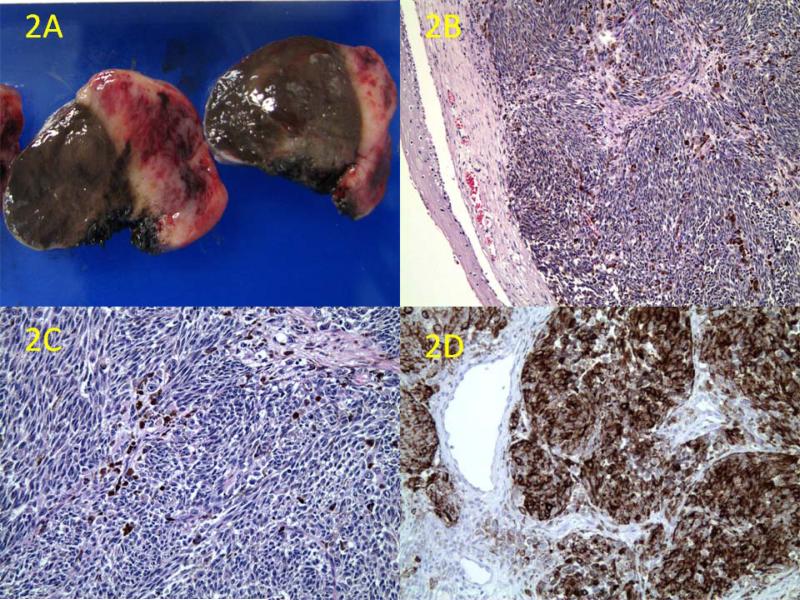
Figure 2.
A: Cross-section examination of the tumor reveals dark-brown pigmentation in the mucosa and tumor parenchyma with areas of hemorrhage and necrosis. B: Microscopic section shows the hypercellular tumor partially covered by squamous mucosa, along with areas of pigmentation (H&E, original magnification ×50). C: The tumor is composed of heavily pigmented pleomorphic spindle cells with mitotic figures (H&E, original magnification ×100). D: Immunohistochemistry shows diffuse positivity for HMB-45 (original magnification ×100)

