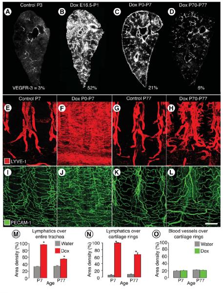
Figure 4. Age-related decrease in severity of VEGF-C driven lymphangiectasia.
A-D, Decreased severity of lymphangiectasia in CCSP-VEGF-C mice on doxycycline beginning at E16.5, P0, or P70. Photographic montages of lungs showing age-related differences in abundance of lymphatics (VEGFR-3 immunoreactivity, white), and corresponding area densities of white pixels (%), in CCSP-VEGF-C mice on water at P3 (A) or on doxycycline from E16.5 to P1 (B), P0 to P7 (C), or P70 to P77 (D). E-L, Lymphatics (E-H, LYVE-1, red) and blood vessels (I-L, PECAM-1, green) in tracheal whole mounts from newborn (E, F, I, J) and adult (G, H, K, L) CCSP-VEGF-C mice on water (E, G, I, K) or doxycycline (F, H, J, L) for 7 days. E, G, Segmented pattern of lymphatics in neonatal and adult mice on water. F, Sheet-like lymphangiectasia in neonatal mouse on doxycycline from P0 to P7. H, Sprouting lymphangiogenesis in adult mouse on doxycycline from P70 to P77. I-L, No apparent differences in blood vessels of neonatal or adult mice on water or doxycycline. M-O, Area density of lymphatics over entire trachea (M), over cartilage rings (N), and corresponding blood vessels over cartilage rings (O). * P < 0.05 vs. water. Scale bar: 450 μm (A, C); 350 μm (B); 600 μm (D); 200 μm (E-L).

