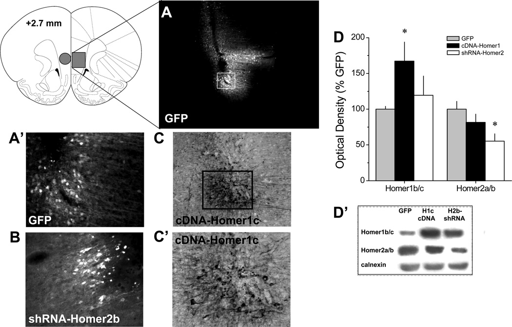
Figure 3. Verification of the in vivo transduction efficacy of the AAVs constructs within vmPFC.
A, Micrograph (4 × magnification of tissue section) of immunofluorescence for the GFP reporter in the vmPFC of Rat #14. This rat exhibited the greatest lateral and dorsal spread of AAV from the microinjector tip of the 30 rats infused with AAV. Despite this, there was little evidence for AAV spread into the dmPFC. A’, 20 × magnification of the immunofluorescence from the GFP reporter on our control AAV from Panel A. B, 20 × magnification of the immunofluorescence from the GFP reporter on shRNA-Homer2b. C, 10× magnification of immunostaining for the HA tag on cDNA-Homer1c, demonstrating the typical pattern of AAV spread from the microinjector tip. C’, 20 × magnification of the immunostaining for the HA Tag from Panel C. D, Summary of the effects of the AAV-GFP (GFP), cDNA-Homer1c or shRNA-Homer2b upon the protein expression of Homer1b/c and Homer2a/b within vmPFC of drug-naïve rats (tissue punch indicated by circle in coronal section of brain). The data represent the mean ± SEM of 5 rats/AAV, expressed as a percentage of GFP controls. *p<0.05 vs. GFP controls (LSD post-hoc tests). D’, Representative immunoblots for Homer1b/c, Homer2a/b and calnexin from AAV-infused animals.

