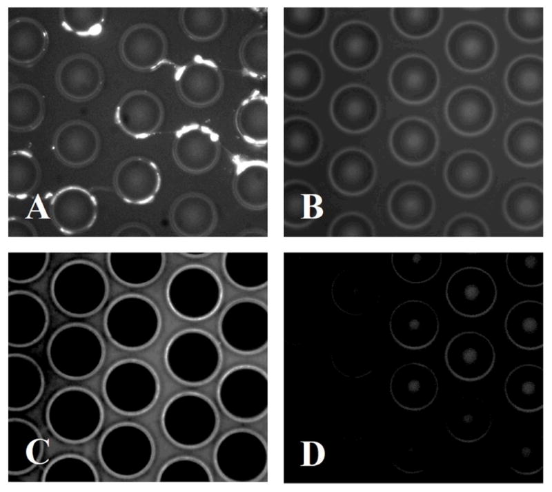
Figure 2.

(A) Fluorescence image of poorly solubilized membrane proteins isolated on the μSPE device. (B) Control image of the μSPE bed incubated with FITC-avidin without first infusing the cell lysate showing minimal nonspecific adsorption of the dye-labeled avidin complex. (C) Fluorescence image of well-solubilized membrane proteins isolated on the μSPE bed. (D) μSPE bed after release of membrane proteins with DTT.
