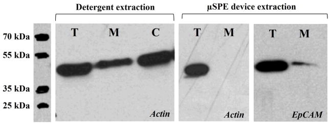
Figure 4.

Actin Western blots demonstrating for detergent-based extraction and the μSPE extraction using actin as the model cytosolic protein. Also shown is the EpCAM Western blot of the membrane protein fraction eluted from the μSPE device to show that there were membrane proteins from the MCF-7 cell lysate in the fraction. For these Western blots, approximately 5 × 10⁶ MCF-7 cells were lysed and taken to a total volume of 1.0 mL. This lysate was either directly loaded onto the gel (30 μL) for Western analysis or diluted ~1000-fold with 100 μL processed using the μSPE device. Due to the limited bed capacity of the μSPE device, the EpCAM band intensity was much weaker for the μSPE device compared to direct processing of the lysate.
