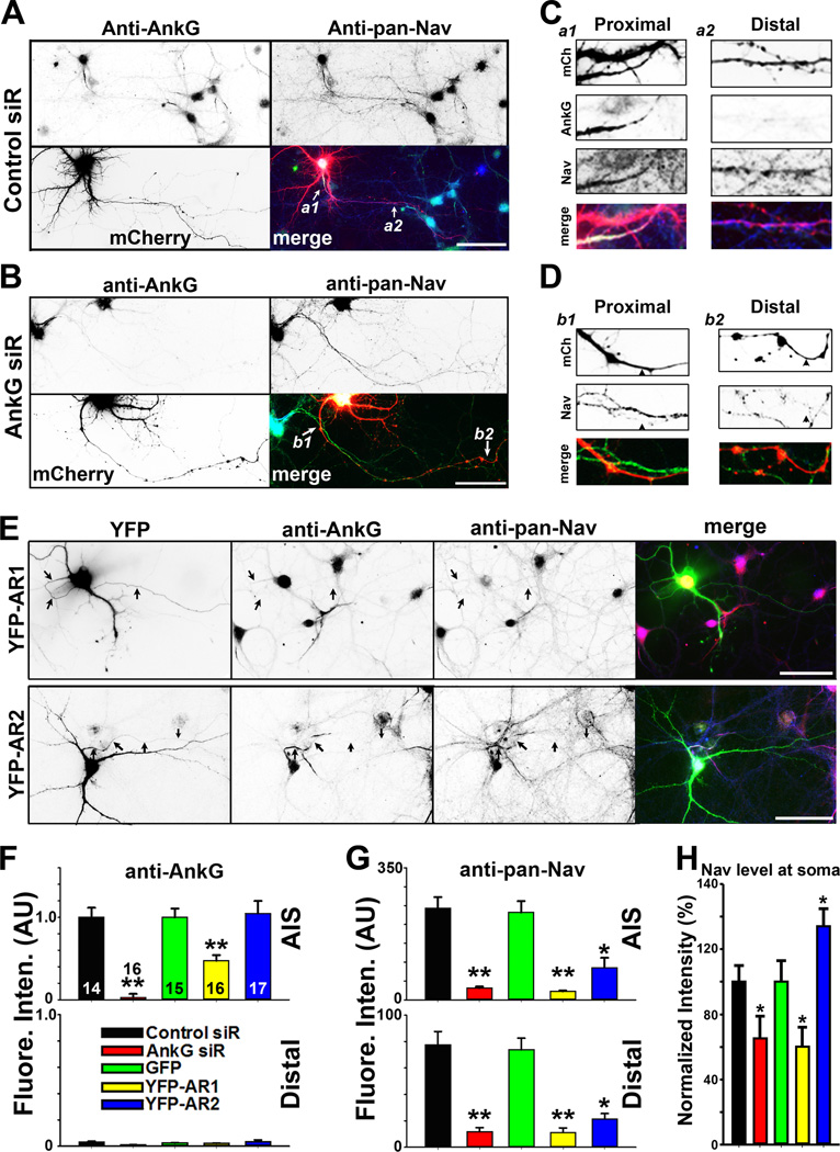
Figure 3. Disrupting AnkG-KIF5 interaction suppresses axonal targeting of Nav channels.
A, Control siRNA had no effect on endogenous AnkG and Nav channels along axons. Hippocampal neurons were transfected with control siRNA at 5 DIV, fixed and stained at 21 DIV. Signals are inverted in gray-scale images. In merged image: anti-AnkG, blue; anti-pan-Nav, green; mCherry from the siRNA vector, red. White arrows, proximal and distal axons. B, Knocking down AnkG with siRNA reduced the overall Nav channel level along the entire axon. C, Two enlarged areas (3 fold) in A. a1, proximal axon; a2, middle-distal axon. D, Two enlarged areas (3 fold) in B. b1, proximal axons; b2, distal axons. Black arrowheads, axons with AnkG siRNA. E, YFP-AR1, but not YFP-AR2, reduced AnkG (red in merged) and Nav channel (blue in merged) levels at the AIS. Hippocampal neurons were transfected at 12 DIV, fixed and stained at 17 DIV. In single images, signals are inverted. Black arrows, proximal axons. Scale bars, 100 µm. F, Summary of the effect of siRNA knock down of endogenous AnkG and over-expression of YFP-AR1 and YFP-AR2 (transfected on 12 DIV and fixed on 17 DIV) on endogenous AnkG along axons. Fluorescence intensities were normalized with Control siRNA or GFP. G, Summary of the effect on endogenous Nav channels along axons. H, Normalized fluorescence intensity of the anti-pan-Nav staining at the soma of the neurons. The "n" number is indicated in each column in the upper panel of F. Unpaired t-test was used for the siRNA experiments and One-Way ANOVA followed by Dunnett's test was used for the experiments of YFP-AR1 and YFPAR2. *, p < 0.05; **, p < 0.01. See also Figure S3.

