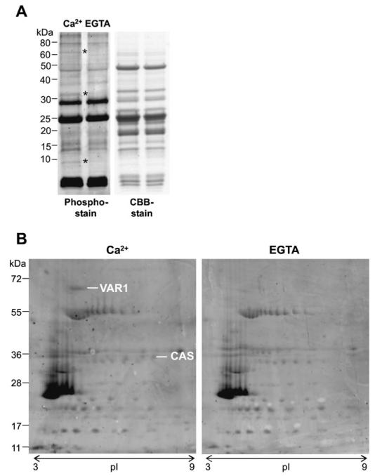
Fig. 1. Calcium-dependent phosphorylation of pea thylakoid proteins.
(A) The left panel displays a Pro-Q Diamond phosphoprotein gel stain that reveals three differentially phosphorylated protein bands (indicated with an asterisk) upon the addition of 1 mM Ca2+, compared with 1 mM EGTA. The right panel is the protein loading control (CBB=Coomassie Brilliant Blue). (B) 2D protein separation according to isoelectric point (pI) and size (kDa) of the same Ca2+-dependent phosphorylation assay followed by a Pro-Q Diamond phosphoprotein gel stain (experiment 1). Two protein spots were identified as the FtsH protease, Variegated 1 (VAR1) and ‘Calcium sensing’ protein (CAS).

