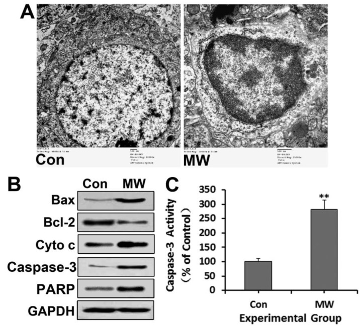
Figure 5.
Neuronal apoptosis of rat hippocampus observed by TEM (A) 6 h after 30 mW/cm2 microwave exposure. All immunoblotting analyses (B), except detection of cytochrome c, were performed using whole hippocampal lysates. After sucrose gradient centrifugation, the supernatant was used as the cytosolic fraction for cytochrome c detection. The immunoblotting results further confirmed the upregulation of Bax, cytochrome c, cleaved caspase-3 and PARP, and the down-regulation of Bcl-2. Caspase-3 activity (C) also increased, as assessed by ELISA. Significant differences are as follows: vs. control, **p < 0.01 (t test). Scale bars: 500 nm.

