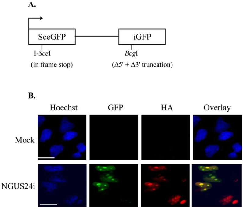
Figure 1.
Efficient expression of I-SceI from an Adenoviral vector based system in T98Gs with an integrated pDRGFP substrate. (A) Schematic diagram of reporter plasmid pDRGFP as described in [12]. (B) Representative IF staining of Ad-infected Clone 10. Cells were seeded onto glass coverslips and infected with Ad expressing I-SceI (NGUS24i). Coverslips were harvested at 72 h post infection. GFP+ cells were scored as HDR competent. Cells were stained with HA-specific Ab to detect HA-I-SceI expressed from NGUS24i. Scale bar = 5 µm for all panels and all figures.

