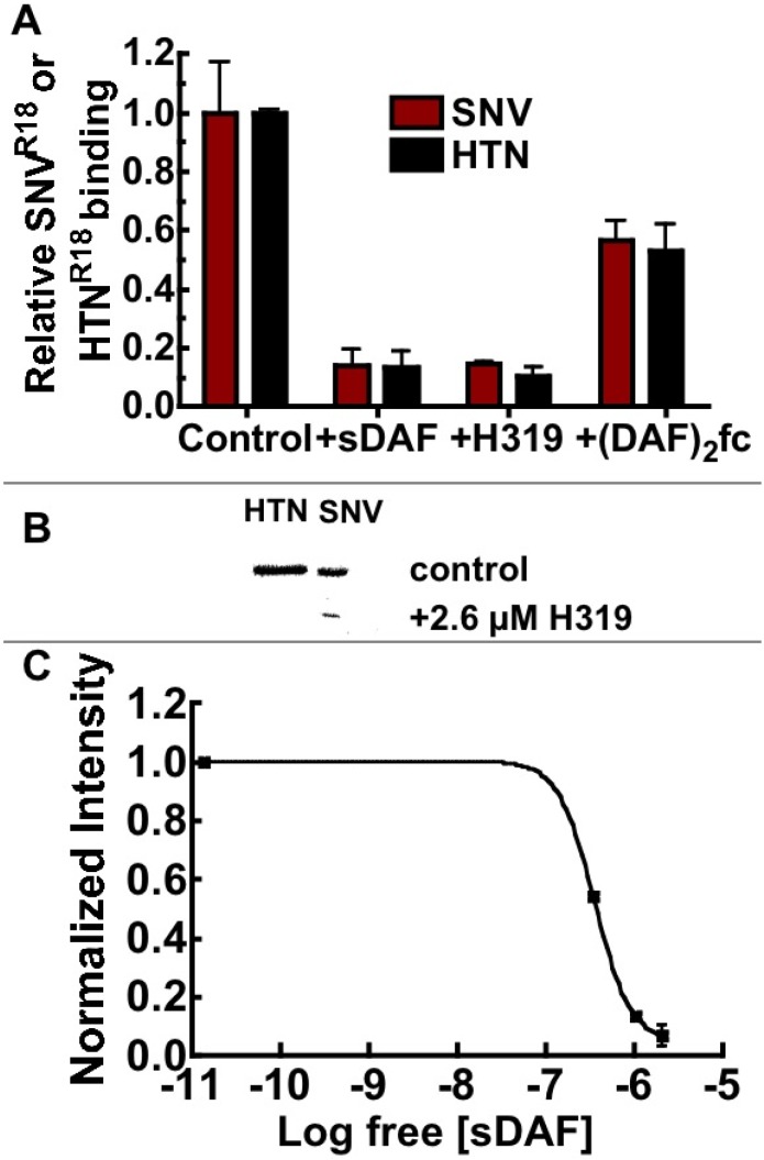
Figure 5.
(A) Soluble DAF (sDAF) is significantly better than (DAF)2-Fc at blocking cell binding and entry of SNV. Flow cytometry analysis of Tanoue B cell-binding inhibition of SNVR18 or HTNR18 using 1.0 µM sDAF, 26 µM H319 polyclonal antibody against DAF and 18.0 µM (DAF)2-Fc. sDAF and H319 inhibited cellular binding of SNV and HTN by >90% compared to <50% associated with (DAF)2-Fc. (B) Western blotting of Vero E6 cells infected with HTN and SNV (control) and cells pretreated with 2.6 µM H319 anti-DAF antibody. (C) Analysis of inhibition of infection of Vero E6 cells by SNV with sDAF, measuring viral N-protein by western blot, which is presented as of a plot of normalized intensity versus concentration of sDAF. Quantitative analysis of the gel was performed using a Biorad Molecular Imager, ChemiDoc XRS+ equipped with Image Lab Software 4.1 [38].

